FIELD: medicine.
SUBSTANCE: invention relates to medicine, namely to oncology, and can be used for detection of tumour cells in surgical material for breast cancer by fluorescent diagnostics. Analysed tissue fragment is stained with an aqueous solution of erythrosine. Flash photolysis is used to record 10 to 30 kinetic curves of delayed fluorescence decay of the coloured sample. Radiation source used is a laser with wavelength of 532 nm with a pulse duration of 15 ns, a pulse repetition frequency of at least 10 Hz with a recording duration of each curve of at least 50 mcs with resolution of 0.1 mcs. Annihilation delayed fluorescence is separated from the total recorded signal by approximating a kinetic decay curve with two exponents. Area under the section of the kinetic curve to the point of intersection of the approximating exponents for each pulse is calculated and normalized to the value of the area for the first pulse. Obtained values are presented as a function of change of integral intensity S(i), where i is the serial number of the pulse, the data S(i) are approximated by a function of the form
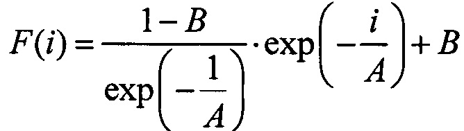 ,
,
where A is the rate of change of the integral intensity during pulsed irradiation, B is the relative decrease in the integral intensity after the last pulse in the series compared to the integral intensity of the annihilation delayed fluorescence after the first pulse. If the value A is more than 9.5 pulse-1 and the value B is more than 0.83, there are no tumour cells in the surgical material. If one or both values are below these values, the presence of tumour cells is detected in the surgical material.
EFFECT: method enables detecting tumour cells in the resection margins and sentinel lymph nodes in breast cancer and determining the indications for axillary lymph node dissection, breast resection and postoperative radiation therapy with high sensitivity and high prognostic value of negative result, due to detection of tumour cells in surgical material for breast cancer by flash photolysis.
1 cl, 9 dwg, 1 tbl, 3 ex
| Title | Year | Author | Number |
|---|---|---|---|
| METHOD OF PREDICTING HIGH RISK OF REGIONAL METASTASIS IN BREAST CANCER | 2023 |
|
RU2816441C1 |
| METHOD OF PREDICTING RESISTANCE TO NEOADJUVANT CHEMOTHERAPY IN PATIENTS WITH BREAST CANCER | 2022 |
|
RU2802671C1 |
| METHOD OF SEARCHING FOR SIGNAL LYMPH NODES IN BREAST CANCER USING ULTRASONIC VISUALIZATION AND INTRADERMAL PARAREOLAR ADMINISTRATION OF SONOVUE ECHOCONTRAST DRUG | 2022 |
|
RU2813026C1 |
| METHOD OF SIGNAL LYMPH NODE BIOPSY IN PATIENTS WITH BREAST CANCER | 2014 |
|
RU2549488C1 |
| SENTINEL LYMPH NODE DIAGNOSTIC METHOD IN STOMACH CANCER | 2007 |
|
RU2354288C1 |
| SENTINEL LYMPH NODES DIAGNOSING METHOD IN GASTRIC CANCER | 2020 |
|
RU2727251C2 |
| METHOD OF INDIVIDUAL PRE-RADIATION PREPARATION OF PATIENTS SUFFERING FROM BREAST CANCER | 2008 |
|
RU2377020C1 |
| METHOD FOR CONSERVATION TREATMENT OF INVASIVE CERVICAL CANCER | 2013 |
|
RU2535614C1 |
| METHOD FOR DETERMINING CALCITONIN AND CANCER-EMBRYO ANTIGEN IN PUNCTATE FOCAL LIVER FORMATION | 2020 |
|
RU2736693C2 |
| METHOD OF DIAGNOSING REGRESSION OF BREAST CANCER AFTER NEOADJUVANT DRUG THERAPY | 2022 |
|
RU2806299C1 |

